SARS-CoV-2 Publications

Nguyen LC, Yang D, Nicolaescu V, Best TJ, Gula H, Saxena D, Gabbard JD, Chen SN, Ohtsuki T, Friesen JB, Drayman N, Mohamed A, Dann C, Silva D, Robinson-Mailman L, Valdespino A, Stock L, Suárez E, Jones KA, Azizi SA, Demarco JK, Severson WE, Anderson CD, Millis JM, Dickinson BC, Tay S, Oakes SA, Pauli GF, Palmer KE, Consortium TNCCC, Meltzer DO, Randall G, Rosner MR. Cannabidiol inhibits SARS-CoV-2 replication through induction of the host ER stress and innate immune responses. Sci Adv. 2022 Jan 20:eabi6110. Epub ahead of print. PMID: 35050692.
El-Shennawy L, Hoffmann AD, Dashzeveg NK, McAndrews KM, Mehl PJ, Cornish D, Yu Z, Tokars VL, Nicolaescu V, Tomatsidou A, Mao C, Felicelli CJ, Tsai CF, Ostiguin C, Jia Y, Li L, Furlong K, Wysocki J, Luo X, Ruivo CF, Batlle D, Hope TJ, Shen Y, Chae YK, Zhang H, LeBleu VS, Shi T, Swaminathan S, Luo Y, Missiakas D, Randall GC, Demonbreun AR, Ison MG, Kalluri R, Fang D, Liu H. Circulating ACE2-expressing extracellular vesicles block broad strains of SARS-CoV-2. Nat Commun. 2022 Jan 20;13(1):405. doi: 10.1038/s41467-021-27893-2. PMID: 35058437; PMCID: PMC8776790.
Neutralization effects of evACE2 on RBD-binding and SARS-CoV-2 variant infections. a Schematic depiction of the cell-based neutralization assay. b Representative flow profiles showing the percentage (fluorescence mean intensity) of RBD-AF647 binding (at 16 and 3.3 nmol/L) to ACE2+ HEK-293 cells, inhibited by rhACE2 and ACE2+ EVs (evACE2) isolated from HEK-293 and HeLa cells (HEK-EV1 and HeLa-EV2, respectively) whereas ACE2− EVs (evCon) had no neutralization effects (no RBD in black, PBS in dark blue, rhACE2 in orange, evCon in light blue, and evACE2 in green). c IC50 of rhACE2 (orange line) and ACE2 in the EVs from ACE2+ HEK (ev1ACE2) and HeLa (ev2ACE2) cells (green lines) on 16 nM RBD-host cell binding (%). GraphPad Prism 9.0.2 was used to calculate the IC50. N = 2 experiments with two technical replicates for each. Data are presented as mean values ± SD. d IC50 of evACE2, ev1 from HEK and ev2 from HeLa cells (green lines), and rhACE2 (orange line) neutralizing infections by wild-type (WT) S+ pseudotyped SARS-CoV-2. GraphPad Prism 9.0.2 was used to calculate the IC50. N = 2 experiments with two technical replicates for each. Data are presented as mean values ± SD. e IC50 (nM) of ACE2 in ev1ACE2 (HEK) (green line) and rhACE2 (orange line) upon wild-type SARS-CoV-2 infection. GraphPad Prism 9.0.2 was used to calculate the IC50 with three biological replicates. Data are presented as mean values ± SD. f Distinct effects of ACE2+ EVs (green lines) and ACE2− control EVs (light blue line) on inhibiting Vero-6 cell death caused by SARS-CoV-2. N = 2 experiments with three biological replicates each. Data are presented as mean values ± SD. g The IC50 of ev1ACE2 (HEK) neutralizing infections by pseudotyped SARS-CoV-2 expressing WT (black), B.1.1.7 (α) variant (red), B1.351 (β) variant (dark blue) and B.1.617.2 (δ) (light green) S protein. GraphPad Prism 9.0.2 was used to calculate the IC50. N = 2 experiments with two technical replicates each. Data are presented as mean values ± SD. h Effects of ev1ACE2 (HEK) on protecting Vero-6 cell viability against infections of SARS-CoV-2 WT (black), B.1.1.7 (α) variant (red) and B1.351 (β) variant (dark blue) (n = 3 biological replicates). Data are presented as mean values ± SD.
A multifunctional neutralizing antibody‐conjugated nanoparticle is developed to capture and inactivate SARS‐CoV‐2. It efficiently captures SARS‐CoV‐2 pseudovirions and completely blocks viral infection of host cells in vitro through the surface neutralizing antibodies. In addition, its photothermal function inactivates viruses upon light irradiation. More importantly, it treats authentic SARS‐CoV‐2 infection in vivo outperforming neutralizing antibodies.

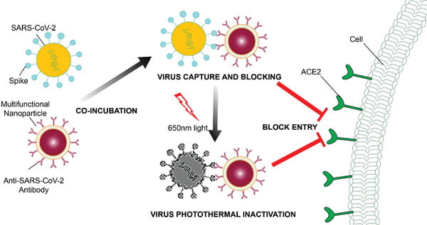
Cai X, Chen M, Prominski A, Lin Y, Ankenbruck N, Rosenberg J, Nguyen M, Shi J, Tomatsidou A, Randall G, Missiakas D, Fung J, Chang EB, Penaloza-MacMaster P, Tian B, Huang J. A Multifunctional Neutralizing Antibody-Conjugated Nanoparticle Inhibits and Inactivates SARS-CoV-2. Adv Sci (Weinh). 2022 Jan;9(2):e2103240. doi: 10.1002/advs.202103240. Epub 2021 Nov 10. PMID: 34761549; PMCID: PMC8646742.

Gray LT, Raczy MM, Briquez PS, Marchell TM, Alpar AT, Wallace RP, Volpatti LR, Sasso MS, Cao S, Nguyen M, Mansurov A, Budina E, Watkins EA, Solanki A, Mitrousis N, Reda JW, Yu SS, Tremain AC, Wang R, Nicolaescu V, Furlong K, Dvorkin S, Manicassamy B, Randall G, Wilson DS, Kwissa M, Swartz MA, Hubbell JA. Generation of potent cellular and humoral immunity against SARS-CoV-2 antigens via conjugation to a polymeric glyco-adjuvant. Biomaterials. 2021 Nov;278:121159. doi: 10.1016/j.biomaterials.2021.121159. Epub 2021 Sep 30. PMID: 34634664; PMCID: PMC8482845.

Volpatti LR, Wallace RP, Cao S, Raczy MM, Wang R, Gray LT, Alpar AT, Briquez PS, Mitrousis N, Marchell TM, Sasso MS, Nguyen M, Mansurov A, Budina E, Solanki A, Watkins EA, Schnorenberg MR, Tremain AC, Reda JW, Nicolaescu V, Furlong K, Dvorkin S, Yu SS, Manicassamy B, LaBelle JL, Tirrell MV, Randall G, Kwissa M, Swartz MA, Hubbell JA. Polymersomes Decorated with the SARS-CoV-2 Spike Protein Receptor-Binding Domain Elicit Robust Humoral and Cellular Immunity. ACS Cent Sci. 2021 Aug 25;7(8):1368-1380. doi: 10.1021/acscentsci.1c00596. Epub 2021 Jul 21. PMID: 34466656; PMCID: PMC8315245.

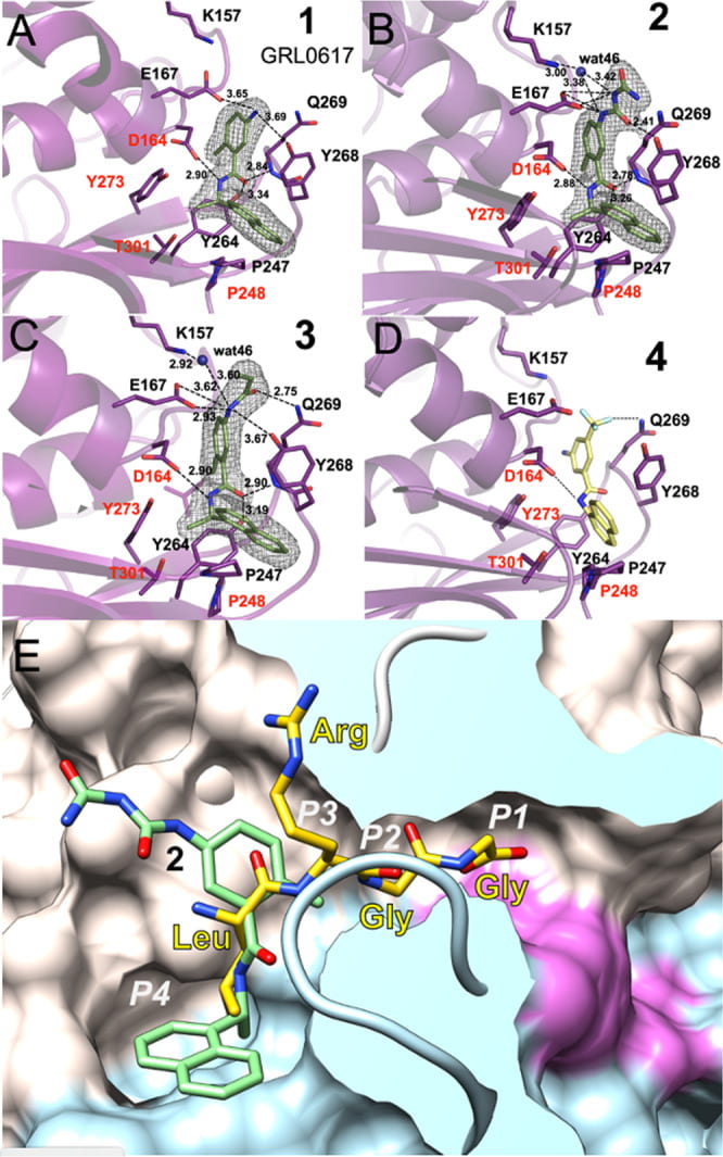
Drayman N, DeMarco JK, Jones KA, Azizi SA, Froggatt HM, Tan K, Maltseva NI, Chen S, Nicolaescu V, Dvorkin S, Furlong K, Kathayat RS, Firpo MR, Mastrodomenico V, Bruce EA, Schmidt MM, Jedrzejczak R, Muñoz-Alía MÁ, Schuster B, Nair V, Han KY, O’Brien A, Tomatsidou A, Meyer B, Vignuzzi M, Missiakas D, Botten JW, Brooke CB, Lee H, Baker SC, Mounce BC, Heaton NS, Severson WE, Palmer KE, Dickinson BC, Joachimiak A, Randall G, Tay S. Masitinib is a broad coronavirus 3CL inhibitor that blocks replication of SARS-CoV-2. Science. 2021 Aug 20;373(6557):931-936. doi: 10.1126/science.abg5827. Epub 2021 Jul 20. PMID: 34285133; PMCID: PMC8809056.

A drug repurposing screen identifies multiple safe-in-human drugs that inhibit OC43 infection. A. Schematic of the screen. A549 cells expressing H2B-mRuby were infected with OC43 (MOI 0.3), treated with drugs, incubated for 4 days at 33°C, and stained for the viral nucleoprotein. B. Screen results showing the %OC43 staining of mock-infected cells (green), no-drug controls (black), drugs with no effect on OC43 infection (blue), and screen hits (red). Overall agreement between the two repeats is high (R2=0.81) C. Dose response curves of remdesivir and the top hits from the screen, n = 3. Individual measurements are shown as semi-transparent circles (note that some circles overlap). Additional dose response curves are shown in Fig. S1.
SARS-CoV-2 enters host cells through its viral spike protein binding to angiotensin-converting enzyme 2 (ACE2) receptors on the host cells. Here, we show that functionalized nanoparticles, termed “Nanotraps,” completely inhibited SARS-CoV-2 infection by blocking the interaction between the spike protein of SARS-CoV-2 and the ACE2 of host cells. The liposomal-based Nanotrap surfaces were functionalized with either recombinant ACE2 proteins or anti-SARS-CoV-2 neutralizing antibodies and phagocytosis-specific phosphatidylserines. The Nanotraps effectively captured SARS-CoV-2 and completely blocked SARS-CoV-2 infection to ACE2-expressing human cell lines and primary lung cells; the phosphatidylserine triggered subsequent phagocytosis of the virus-bound, biodegradable Nanotraps by macrophages, leading to the clearance of pseudotyped and authentic virus in vitro. Furthermore, the Nanotraps demonstrated an excellent biosafety profile in vitro and in vivo. Finally, the Nanotraps inhibited pseudotyped SARS-CoV-2 infection in live human lungs in an ex vivo lung perfusion system. In summary, Nanotraps represent a new nanomedicine for the inhibition of SARS-CoV-2 infection.

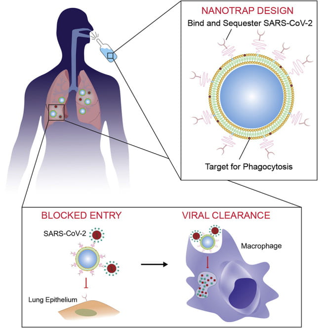
Chen M, Rosenberg J, Cai X, Hsuan Lee AC, Shi J, Nguyen M, Wignakumar T, Mirle V, Edobor AJ, Fung J, Donington JS, Shanmugarajah K, Lin Y, Chang E, Randall G, Penaloza-MacMaster P, Tian B, Madariaga ML, Huang J. Nanotraps for the containment and clearance of SARS-CoV-2. Matter. 2021 Jun 2;4(6):2059-2082. doi: 10.1016/j.matt.2021.04.005. Epub 2021 Apr 22. PMID: 33907732; PMCID: PMC8062026.

Teplensky MH, Distler ME, Kusmierz CD, Evangelopoulos M, Gula H, Elli D, Tomatsidou A, Nicolaescu V, Gelarden I, Yeldandi A, Batlle D, Missiakas D, Mirkin CA. Spherical nucleic acids as an infectious disease vaccine platform. Proc Natl Acad Sci U S A. 2022 Apr 5;119(14):e2119093119. doi: 10.1073/pnas.2119093119. Epub 2022 Mar 21. PMID: 35312341.

Nguyen LC, Yang D, Nicolaescu V, Best TJ, Ohtsuki T, Chen SN, Friesen JB, Drayman N, Mohamed A, Dann C, Silva D, Gula H, Jones KA, Millis JM, Dickinson BC, Tay S, Oakes SA, Pauli GF, Meltzer DO, Randall G, Rosner MR. Cannabidiol Inhibits SARS-CoV-2 Replication and Promotes the Host Innate Immune Response. bioRxiv [Preprint]. 2021 Mar 10:2021.03.10.432967. doi: 10.1101/2021.03.10.432967. PMID: 33758843; PMCID: PMC7987002.

Structure of SARS-CoV-2 hexamer with the bound Tipiracil in surface representation. Tipiracil bound to each subunit active site is shown with all atoms in color spheres (carbon, chlorine, nitrogen, oxygen in white, green, blue, and red, respectively).
Kim Y, Wower J, Maltseva N, Chang C, Jedrzejczak R, Wilamowski M, Kang S, Nicolaescu V, Randall G, Michalska K, Joachimiak A. Tipiracil binds to uridine site and inhibits Nsp15 endoribonuclease NendoU from SARS-CoV-2. Commun Biol. 2021 Feb 9;4(1):193. doi: 10.1038/s42003-021-01735-9. PMID: 33564093; PMCID: PMC7873276.
Osipiuk J, Azizi SA, Dvorkin S, Endres M, Jedrzejczak R, Jones KA, Kang S, Kathayat RS, Kim Y, Lisnyak VG, Maki SL, Nicolaescu V, Taylor CA, Tesar C, Zhang YA, Zhou Z, Randall G, Michalska K, Snyder SA, Dickinson BC, Joachimiak A. Structure of papain-like protease from SARS-CoV-2 and its complexes with non-covalent inhibitors. Nat Commun. 2021 Feb 2;12(1):743. doi: 10.1038/s41467-021-21060-3. PMID: 33531496; PMCID: PMC7854729.
Ligands binding to SARS-CoV-2 PLpro. A Compound 1 binding to PLpro. B Compound 2 binding to PLpro. C Compound 3 binding to PLpro. D Model of compound 4 (yellow sticks) binding to PLpro. Ligands are shown as green sticks and PLpro is in magenta. Dashed lines show hydrogen bonds, water molecules are shown as blue spheres. In A–C the 2Fo − mFc electron density maps are shown as a grey mesh, contoured at 1.2 σ. E Compound 2 (green sticks) binds to a groove on the surface of PLpro protein (surface of palm subdomain is in white and thumb subdomain is in light blue) with the active site catalytic triad surface is shown in red in the end of a slender tunnel. Peptide LRGG from ubiquitin structure in complex with SARS PLpro (PDB id: 4MOW) is shown in yellow and peptide positions corresponding P1–P4 sites are marked in white.